
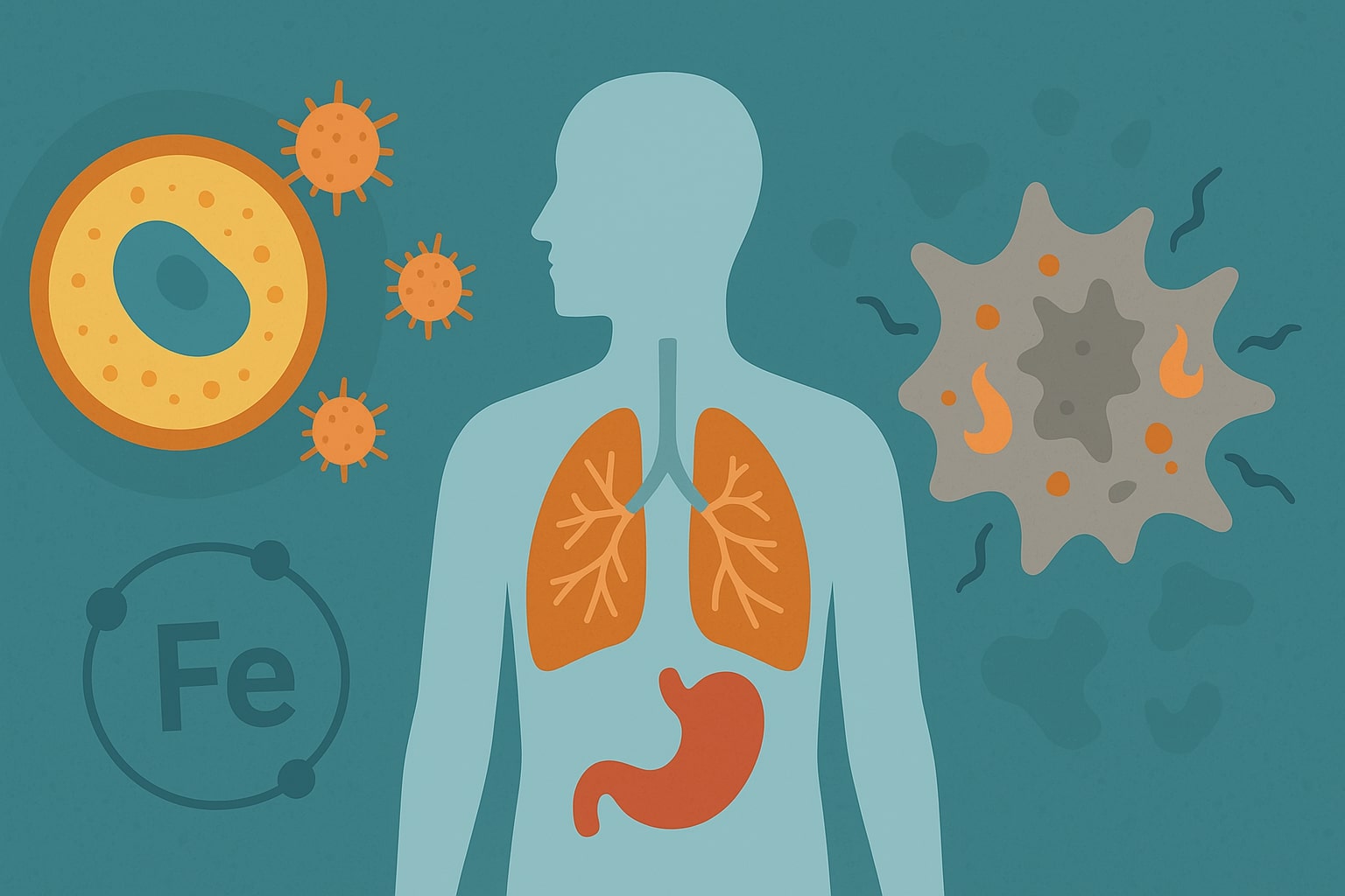
Cells are designed to die in ways that serve a purpose. Not all cell death is chaotic or harmful. In fact, some forms are tightly regulated and essential to health. One of the more recently identified types is ferroptosis, a process distinct from better-known forms like apoptosis or necrosis. Researchers have discovered that ferroptosis plays a role in several important areas, including cancer, brain health, and immune function.
There’s also growing interest in how nutrients and metabolites may affect this process. One example is C15, or pentadecanoic acid. This fatty acid has been studied for its potential to influence inflammation and cellular metabolism, both of which intersect with ferroptosis in emerging research.
Today, we’re diving into ferroptosis so that you can empower yourself and improve your health.
Defining Ferroptosis
Ferroptosis is a form of regulated cell death driven by the accumulation of iron and lipid-based reactive oxygen species. What sets it apart from other types of cell death is its dependence on iron and oxidative damage to the cell’s lipid membranes. Unlike apoptosis, which involves DNA fragmentation and cell shrinkage, ferroptosis primarily affects the integrity of the cell membrane through lipid peroxidation.
This process becomes dangerous when the cell can no longer control the buildup of these reactive compounds. Under normal conditions, an enzyme called glutathione peroxidase 4 (GPX4) helps prevent damage by neutralizing lipid peroxides. When GPX4 is impaired or overwhelmed, lipid peroxides accumulate and trigger ferroptosis.
Ferroptosis is not a random event. It’s a carefully orchestrated process with defined molecular pathways that make it both a useful biological mechanism and a potential target for therapeutic intervention.
What Triggers Ferroptosis?
Several factors can trigger ferroptosis, all centering around oxidative stress and iron imbalance. One of the primary initiators is the depletion of glutathione, an antioxidant that cells rely on to detoxify harmful molecules. Without enough glutathione, the activity of GPX4 is reduced, allowing lipid peroxides to build up and damage the cell membrane.
Iron overload is another major factor. Excess intracellular iron can participate in Fenton reactions (chemical processes that generate highly reactive hydroxyl radicals). These radicals damage membrane lipids and accelerate the onset of ferroptosis.
Other contributing triggers include certain chemotherapy drugs, environmental stressors, and disruptions in lipid metabolism. These conditions collectively push the cell toward a state where it can no longer maintain membrane integrity, leading to regulated but irreversible cell death.
Ferroptosis in Disease Research
Ferroptosis has gained attention in medical research due to its involvement in several major diseases. When it comes to cancer, certain tumor cells are especially vulnerable to ferroptosis. Researchers are exploring ways to trigger this process selectively to destroy cancer cells that resist other treatments. Some experimental therapies focus on inhibiting GPX4 or increasing iron levels to induce ferroptosis in tumors.
In neurodegenerative diseases like Alzheimer’s and Parkinson’s, ferroptosis may contribute to neuronal loss. These conditions often involve oxidative stress and iron accumulation in the brain, two key elements of the ferroptotic pathway.
Also, ischemia-reperfusion injury, which occurs when blood supply returns to tissue after a blockage (such as during a heart attack or stroke), can activate ferroptosis due to sudden oxidative stress.
Nutrients, Metabolism, and Ferroptosis
The connection between ferroptosis and metabolism is another area of growing interest. Cells that rely heavily on polyunsaturated fatty acids (PUFAs) in their membranes are more susceptible to ferroptosis because these fats are highly prone to oxidative damage. Enzymes involved in fatty acid metabolism can influence how vulnerable a cell is to lipid peroxidation.
Nutritional components, including certain fatty acids and micronutrients, may modulate ferroptosis sensitivity. For example, selenium plays a role in supporting GPX4 function. Compounds that affect inflammation, antioxidant defenses, or membrane composition are also under investigation for their impact on ferroptosis pathways.
This area of study suggests that diet and supplements could one day play a supportive role in managing diseases associated with ferroptosis, though more research is needed to define their specific effects and applications.
How Ferroptosis Is Studied and Measured
Studying ferroptosis requires a combination of biochemical tools and molecular techniques. Researchers use chemical agents like erastin and RSL3 to induce ferroptosis in cell cultures. These compounds help mimic the conditions that lead to lipid peroxidation and glutathione depletion.
Detection methods focus on measuring lipid ROS (reactive oxygen species), GPX4 activity, intracellular iron levels, and changes in membrane integrity. Advanced imaging, genetic knockdown models, and specific fluorescent probes also support detailed investigations.
Animal models and organoid systems allow scientists to examine how ferroptosis operates in complex biological settings, providing valuable insights into disease mechanisms and therapeutic potential.
Ferroptosis: Where Cell Death Meets Opportunity
Ferroptosis is a highly regulated and biologically significant process with far-reaching implications. From cancer research to brain health and metabolic regulation, understanding ferroptosis may unlock new pathways for treatment and prevention.
While much remains to be explored, especially around the role of diet and natural compounds, the progress so far is promising. As science uncovers more about how ferroptosis is triggered, controlled, and prevented, it becomes increasingly clear that this form of cell death may be key to protecting life itself.